


Introduction
Environmental toxicants are chemicals found in the environment which are harmful to humans and wildlife. Many chemicals used by humans are dispersed in the ecosystem. Mechanisms by which environmental toxicants impart toxicity include formation of free radical and alteration of cellular components [1]. Polychlorinated biphenyls (PCBs) are organic pollutants and are very stable in nature. Since the 1970s, when restrictions were imposed on the production of PCBs, concentrations in environment have shown a gradual decrease. But residues of PCBs still persist in air, soil, water, and sediment and can be detected in biologic tissues in most residents of industrialized countries [2,3
4]. Animal experiments have evidences of the acute effects of PCBs on the liver, kidney, and central nervous system upon oral exposure [5]. The toxicity of PCBs is fairly well described, and embraces changes in neurotransmitter levels and its reuptake, calcium homeostasis, intracellular signaling pathway activation, and for some congeners, it is found to cause cancer [6,7]. The neurotoxicity of PCBs is known to arise due to its ability to induce oxidative stress in the brain and also induce formation of reactive oxygen species (ROS) as PCBs are reduced by cytochrome P450 in to products such as semiquinone and hydroquinone [8]. The neurological impact of PCBs have been comprehensively studied in various animal models of PCB exposure, either as single congeners or mixtures, during different periods of the animal's lifespan [9]. Many of these studies were conducted only on young animals or animals exposed prenatally to PCBs [10,11]. However, a study conducted on adult rats found that spatial learning behavior was altered due to PCB exposure, thereby indicating that PCBs can alter the nervous system in adult animals as well [12,13]. Coenzyme Q10 (CoQ10) is a ubiquinone, which is a fat-soluble vitamin-like substance found in bodies every cell and functions as a coenzyme for a number of key enzymatic steps in the generation of energy inside the cell. CoQ10 has a wide variety of functions and applications in the body. It is the coenzyme for mitochondrial enzymes (complexes I, II and III) as well as enzymes in other parts of the cell. Mitochondrial enzymes of the oxidative phosphorylation pathway are crucial for the production of the high-energy phosphate, adenosine triphosphate (ATP), which is the main currency for cellular function [14,15]. CoQ10 per se or in combination with other drug may be valuable in the cure of numerous health problems, mainly cardiac diseases, breast cancer, diabetes mellitus, immune deficiency, muscular dystrophy, and periodontal disease [16]. Decreased levels of CoQ10 has been noted in several diseases both in animals and humans. Especially in diseases linked to oxidative stress like Alzheimer's disease, diabetes, prion disease and carcinogenesis, CoQ10 biosynthesis is increased [17]. Both genetic and environmental mediators contribute in triggering the Parkinson's disease causing energy pathway impairment and oxidative stress. CoQ10 acting as a mitochondrial nutrient and plays an important role in mitigating both these processes [18]. Also there are strong evidences for the absorption and distribution of CoQ10 to neural tissues as well as preliminary evidences for its role as a neuroprotective in animal models [19].
The important findings from the animal literature as well as the epidemiological studies in humans suggest that the minimum exposure of PCBs is sensitive enough to cause behavioral deficit [9,20]. In addition, results from studies carried out in rodents and monkeys shed light on neurotoxic potentials of PCBs [21,22]. Hence, in the light of existing knowledge, the present study was planned to study the impact of CoQ10 on the oxidative stress induced by oral 28 days exposure of aroclor 1254, a PCB mixture in the brains of Swiss albino mice. For this we estimated the activities of the enzymes superoxide dismutase (SOD), catalase (CAT), glutathione peroxidase (GPx), acetyl cholinesterase (AChE) and levels of lipid peroxidation (LPO) and reduced Glutathione (GSH) in the brains of mice to shed light on the possible mechanism, along with histopathological investigations. L-deprenyl was used as the reference standard as it possesses the property to increase survival of neurons [23].
Materials and Methods
Drugs and Chemicals
All the chemicals used for biochemical assays were obtained from S.D. Fine Chem Ltd. (Mumbai, India), Thomas Baker Chemicals Pvt. Ltd. (Mumbai, India), Hi-media Laboratories Pvt. Ltd. (Mumbai, India). Aroclor 1254 was obtained from Sigma Aldrich Chemicals (St. Louis, MO, USA), CoQ10 was procured and authenticated from M/s. Sterling Biotech Ltd. (Vadodara, India). The standard used for comparison selegiline HCl (L-deprenyl) was obtained from Cipla Ltd. (Mumbai, India). All chemicals used were of analytical grade.
Animals and Treatments
Twenty four female Swiss albino mice (20-30 g) were used in the study. Animals were housed in standard polypropylene cages with wire mesh top and maintained at 23±2℃ and relative humidity 60±5% with 12 hours light-dark cycle. Animals were fed with commercially available standard rodent pellet diet (Amrut rat and mice feed manufactured by Navi Maharashtra Chakan Oil Mill Ltd., Mumbai, India). Water was provided to the animals ad libitum. Experimental procedures were approved by the Institutional Animal Ethics Committee of Bombay College of Pharmacy. The aroclor 1254 (PCBs) was dispersed in corn oil, while CoQ10 was dissolved in 25 µL corn oil. Standard drug L-deprenyl was dissolved in 0.9% saline. Aroclor 1254 was given orally and CoQ10 and L-deprenyl were administered intraparitoneally. Mice were randomized into four groups. The group 1 consisted of vehicle (corn oil, oral) control animals, group 2 was of animals exposed to aroclor 1254 (5 mg/kg, oral), group 3 consisted of animals treated with CoQ10 (10 mg/kg, intraperitoneal injection [IP]) daily followed by aroclor 1254 (5 mg/kg, oral) and group 4 was of L-deprenyl (1 mg/kg, IP) daily followed by aroclor 1254 (5 mg/kg, oral). This administration was done for a period of 28 days. The last dose was administered 24 hours before euthanasia.
Preparation of Homogenate
After 28 days, experimental and control animals were euthanized in CO2 chamber. Brains were immediately taken out and washed thoroughly with ice-cold saline to remove blood. Whole brain was homogenized in 50 mM phosphate buffer pH 7.4 with homogenizer fitted with Teflon plunger. 10% tissue homogenate (w/v) was made. Supernatant was made by centrifuging homogenate at 10,000 rpm for 20 minutes. Homogenate were used for the estimation of LPO and GSH, while supernatant were used for the assay of CAT, SOD, GPx, and AChE activities.
Lipid Peroxidation
The brain tissue homogenate (0.1 mL) was mixed with acetic acid (20%, 1.5 mL), sodium lauryl sulfate (0.2 mL) and thiobarbituric acid (1.5 mL). After incubating at 95℃ for 1 hour the mixture was cooled and 5 mL of n-butanol and pyridine (15:1) were added, shaken vigorously and centrifuged. The absorbance of the homogenate was read at 532 nm against the blank containing all reagents except the homogenate. The inhibition of LPO was determined by the procedure previously described [24]. The values were expressed as nmol of malondialdehyde/mg of protein.
Reduced Glutathione
To 250 µL of brain tissue homogenate, 0.5 mL of 5% trichloroethanoic acid in 1 mM ethylenediaminetetraacetic acid (EDTA) was added. The sample was centrifuged at 2,000 g for 10 minutes. The supernatant was mixed with 2.5 mL of 0.1 M phosphate buffer (pH 8.0) and 100 µL of (0.001%) 5, 5'-dithiobis-2-nitrobenzoic acid (DTNB). Absorbance was read at 412 nm. Values were calculated from a standard graph of GSH treated with same reagent [25]. The amount of reduced glutathione was expressed as µg of GSH/mg protein.
Catalase Aactivity
The procedure followed was as discussed previously [26]. The reaction mixture consisted of 2 mL phosphate buffer (pH 7.0), 0.95 mL of hydrogen peroxide (0.019 M) and 0.05 mL of supernatant in a final volume of 3 mL. Absorbance were recorded at 240 nm every 10 seconds for 1 minute. The results were expressed as units of CAT activity/mg protein.
Superoxide Dismutase Activity
The reaction mixture consisted of 0.05 mL of supernatant, 2 mL of carbonate buffer and 0.5 mL of EDTA solution. The reaction was initiated by the addition of 0.5 mL of epinephrine and the autoxidation of adrenaline to adrenochrome at pH 10.2 was measured by following the change in optical density at 480 nm. Change in optical density every minute was noted at 480 nm against reagent blank. The results were expressed as units of SOD activity/mg protein [27].
Glutathione Peroxidase Activity
The reaction mixture contained 0.5 mL of phosphate buffer (20 mM, pH 7.0), 0.1 mL of glutathione reductase (0.24 U), and 0.1 mL of 10 mM GSH and 0.05 mL of supernatant. Sodium azide (1 mM) was added to the reaction mixture in order to inhibit remnant CAT activity. Thereafter 0.1 mL nicotinamide adenine dinucleotide phosphate-oxidase (NADPH) solution (0.118 mM) was added and the hydroperoxides independent consumption of NADPH was monitored for about 3 minutes. Addition of 0.1 mL hydrogen peroxide (12.5 mM) started the overall reaction and the decrease in absorption at 240 nm was monitored for 5 minutes. The results were expressed as units of GPx activity/mg protein [28].
Acetylcholinesterase Activity
To a cuvette containing 2.6 mL of phosphate buffer (pH 7.5, 0.1 M), 0.4 mL supernatant was added. 100 µL of DTNB reagent was added. The absorbance was measured at 412 nm, when it stopped increasing, absorbance was set to zero. 20 µL of acetylthiocholine was added. Changes in absorbance were recorded for 3 minutes and the change in absorbance per min was calculated. The results were expressed as AChE nmol per minute per g tissue of brain [29].
Histopathological Evaluations
The cerebral cortex was fixed in 10% formalin. The specimens were then processed by standard procedure. The blocks were then sectioned according to hematoxylin and eosin method [30]. Histological sections of five micrometer were taken. The sections were examined under the light microscope and photographed at 100× using Motic camera (BA310; Hong Kong).
Results
Lipid Peroxidation
The malondialdehyde levels, an indicator of LPO were increased in the brain of mice treated with aroclor 1254 (1.85 nmol/mg protein as compared to normal group 0.68 nmol/mg protein). This increase was abated by 10 mg/kg IP dose of CoQ10 (0.55 nmol/mg protein as compared to that of L-deprenyl 0.64 nmol/mg protein) (Table 1).
Reduced Glutathione
Reduced glutathione, a thiol plays a vital role in protecting the cells during endogenous and exogenous free radicals and oxidants mediated insults. The brain GSH levels in mice on 28 days exposure to aroclor 1254 were decreased to 0.25 µg/mg protein from control levels of 0.76 mg/mg protein. Intraperitoneal administration of CoQ10 (10 mg/kg) increased the levels of GSH to 0.42 µg/mg protein. The increase in GSH content by CoQ10 was comparable to the increase obtained by L-deprenyl (0.52 µg/mg protein) (Table 1).
Superoxide Dismutase Activity
SOD is a measure of the oxidative stress produced in the organ as it dismutase O2 anions. Decrease in its activity directly corresponds to the increase in oxidative stress. The significant reduction of 4.43 units/mg protein seen in the aroclor 1254 exposed group as contrast to the control group (8.74 units/mg protein) CoQ10 (10 mg/kg) intraperitoneal administration increased the levels of SOD (5.90 units/mg protein) as compared to that of L-deprenyl (6.73 units/mg protein) (Table 1).
Catalase Activity
The ability of catalase to induce reduction in levels of hydrogen peroxidase was evaluated. Hydrogen peroxidase gives rise to free radicals which are congeners of oxidative stress in our body. The exposure of aroclor 1254 for 28 days resulted in a significant decrease in catalase activity to 0.98 units/mg protein. Supplementation with intraperitoneal CoQ10 increases this activity to 2.21 units/mg protein, which is comparable to L-deprenyl treated group (3.23 units/mg protein) (Table 1).
Glutathione Peroxidase Activity
GPx is mainly responsible for alleviating the effects of free radicals in brain. The decrease in its activity in aroclor 1254 exposed group 3.82 units/mg protein as compared to vehicle control group (19.23 units/mg protein) bears testimony to the oxidative stress inducing effects of aroclor 1254. Co administration of CoQ10 moderately prevented the dramatic decrease in activity (6.98 units/mg protein). The L-deprenyl group also showed an improvement in the enzyme activity (10.70 units/mg protein) (Table 1).
Acetylcholinesterase Activity
Acetylcholinesterase is the main enzyme in brain causing degradation of acetylcholine, which is involved in many motor coordination skills and also memory. The AChE activity of vehicle control group was (230 nmol/min/tissue). The decrease in activity of acetyl cholinesterase (136 nmol/min/tissue) on exposure to aroclor 1254 for 28 days was alleviated on treatment with intraperitoneal CoQ10 treatment (194 nmol/min/tissue). This value bears same level of significance as that obtained with the administration of standard L-deprenyl (219 nmol/min/g tissue) (Table 1).
Histopathological Evaluation
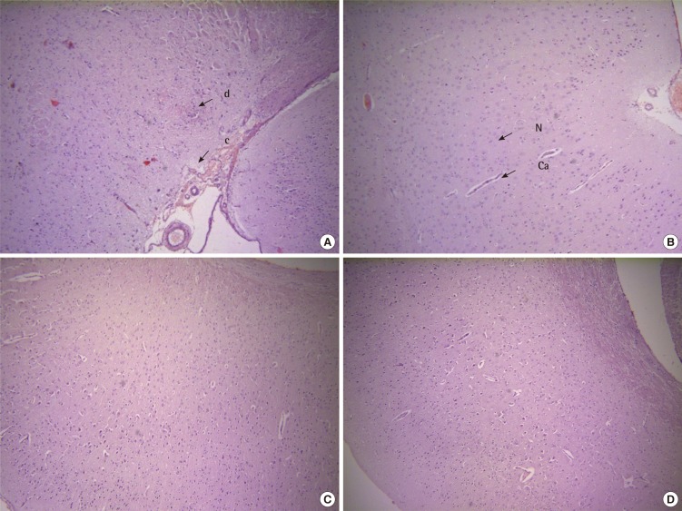
Histopathological evaluation of sections of cerebral cortex of mice brains exposes degeneration, congestion and infiltration in aroclor 1254 exposed group (Figure 1A), as compared to vehicle control group with normal morphology (Figure 1B). Significant prevention of aroclor exposed structural derangements in the cerebral cortex was seen in CoQ10 treated group (Figure 1C) which was comparable to the standard L-deprenyl treated group (Figure 1D).
Discussion
In relation to other organs of the body, the brain is predominantly susceptible to oxidative damage as it undergoes aerobic metabolism and use 20% of the oxygen, generating large amount of ATP molecules [31,32]. It also contains relatively high amount of polyunsaturated fatty acids which are easily oxidizable. Moreover owing to the lipophilic nature of the environmental toxicants, they gain easy access to the brain and hence brain becomes more vulnerable to their toxicity. PCBs form reactive metabolites PCB-epoxides and PCB-quinones. These metabolites are capable of binding to the DNA and RNA which are implicated in the neurodegenerative diseases. PCB induced detrimental effects are associated with the generation of free radicals [33]. Previous evidences suggest that dopaminergic neurons are particularly vulnerable to oxidative stress due to production of ROS due to inherent auto oxidation of dopamine to form H2O2 superoxide anion and dopamine quinines [34,35]. These oxygen radicals can react with cellular nucleophiles, such as GSH. Oral administrations of PCB to rats and primates have been shown to decrease dopamine levels in the mammalian brains in vivo [21]. While a study unraveled that, 52 weeks of exposure to several aroclor mixtures at a high dose in the diet was unsuccessful in producing any functional or morphologic changes indicative of PCB-induced neurotoxicity [36]. On the contrary, the current study shows that sub chronic exposure of aroclor 1254 in mice produced significant neurotoxicity. Many previous studies investigated and reported the role of CoQ10 as neuronal protectant against ROS induced damage and apoptotic cell death. CoQ10 possibly acted through mitochondrial membrane stabilization, when neuronal cells are subjected to oxidative stress [18]. In the present study we investigated the role of CoQ10 in alleviating the oxidative stress induced by oral exposure to aroclor 1254 for 28 days in the brains of Swiss albino mice. The oxidative stress and the free radical generated by the biphenyls are due to the formation of the LPO products leading to the aberrations of neuronal membrane structure. The decrease in activities of radical scavenging enzymes like SOD, CAT, and GPx and levels of GSH resulted in increased oxidative stress in brain. These mechanisms contribute to the PCBs induced neurotoxicity. This investigation revealed a significant decrease in activities of SOD, CAT, GPx and the GSH levels, while a complementary increase in lipid peroxidation was observed on sub acute exposure to aroclor 1254. CoQ10 (5 mg/kg, IP, for 28 days) treatment significantly reversed the detrimental oxidative changes in brain induced by PCBs exposure.
Acetylcholine is known to play an important role in motor coordination and its altered levels are implicated in many neurodegenerative disorders. AChE influences cholinergic transmission and it is known that its activity is inhibited by free radical formation [37]. Motor coordination and cognition is primarily mediated through cholinergic transmission. It is reported that PCB exposure impacts cholinergic system in experimental animals [38]. Our study revealed that intraperitoneal administration of CoQ10 (5 mg/kg, 28 days) treatment significantly abated the changes in anticholinesterase activity induced by aroclor 1254 exposure. Earlier reports have indicated that L-deprenyl indirectly inhibits AChE, but the underlying mechanism is unclear [39]. In the present study L-deprenyl restored the activity of AChE to normal levels. Further, the histopathological evidences of the cerebral cortex of mice brains corroborate the protection offered by CoQ10 in countering the neurotoxicity of PCBs.
We conclude that aroclor 1254 induced oxidative stress mediated neurotoxicity. CoQ10 restored the levels of GSH and the activities of SOD, CAT, GPx, AChE that was adversely wavered by the PCB mixture aroclor 1254 and prevented LPO in the brain region. The treatment outcome obtained is comparable with that of L-deprenyl. These results depict the neuroprotective action of CoQ10 against aroclor 1254 induced brain toxicity.